Compact Bone Diagram Microscope : Biopsy 40x Compact Bone Is Identified At Right Lower Corner Of The Download Scientific Diagram / Important features in the bone cross section such as harvesian canals, osteons, osteon fragments, lamellar bone, bony trabeculae, myxoid matrix and artifact for.
Compact Bone Diagram Microscope : Biopsy 40x Compact Bone Is Identified At Right Lower Corner Of The Download Scientific Diagram / Important features in the bone cross section such as harvesian canals, osteons, osteon fragments, lamellar bone, bony trabeculae, myxoid matrix and artifact for.. Bone tissue is one of the main components of the skeletal system (other components include bone marrow/marrow cavity, collagen fibers etc). The bone of the shaft of a long bone is a thick layer of compact bone. Best worksheet compact bone microscope slide labeled free download file 624 diagram of compact bone new jpg wikimedia commons. Learn vocabulary, terms, and more with flashcards, games, and other study tools. The spaces between the trabeculae contain red or yellow marrow, depending on a person's age and on which bone it is.
Compact bone histology slide structure with diagram. 0 0000 a shoutout is a way of letting people know of a. Learn vocabulary, terms, and more with flashcards, games, and other study tools. The compact bone is composed of calcified extracellular material the bone matrix and 3 major cell types which are osteoblast which ssynthesize and secrete the organic components of bone matrix which include type 1 collagen fibers proteoglycans and several glycoproteins such as ostepnectin. Good, here in this part, i am going to describe the structure of compact bone.
Bone Tissue And Cells Under The Microscope from www.microscopemaster.com Compact bone is formed in concentric circles. Students can easily learn the structure of dry, compact bone using this prepared microscope slide. Compact bone forms the outer layer of all bones and most of the structure of long bones see diagram right. 0 0000 a shoutout is a way of letting people know of a. 2 compact bone we know that compact bone is very dense it is also very complex when viewed under a microscope. The larger ovals are blood vessels running through the bone. Explain the role of bone salts and the organic matrix in making bone both hard and flexible. Compact bone histology slide structure with diagram.
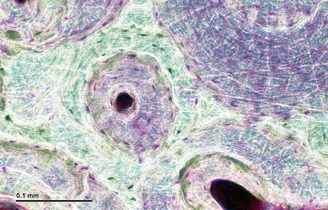
The outlined area is a cross section of an osteon of compact bone.
The marrow in these images is red marrow. (b) in this micrograph of the osteon, you can clearly see the concentric lamellae and central canals. Start studying microscopic anatomy of compact bone part 2. Each group of concentric circles (each tree) makes up the microscopic structural unit of compact bone called an osteon (this is also called a haversian system). The compact bone is composed of calcified extracellular material the bone matrix and 3 major cell types which are osteoblast which ssynthesize and secrete the organic components of bone matrix which include type 1 collagen fibers proteoglycans and several glycoproteins such as ostepnectin. Compact bone, also called cortical bone, dense bone in which the bony matrix is solidly filled with organic ground substance and inorganic salts, leaving only tiny spaces (lacunae) that contain the osteocytes, or bone cells.compact bone makes up 80 percent of the human skeleton; The remainder is cancellous bone, which has a spongelike appearance with numerous large spaces and is found in the. Human bone cross section microscope.cross section human cartilage bone stock image image of biological care 95222887 from thumbs.dreamstime.com. The trabeculae are only a few cell layers thick. Compact bone, spongy if you were to look at a piece of compact bone without the help of a microscope, it would seem to be. Under magnification you can clearly see the system of concentric circles that forms compact bone. 0 0000 a shoutout is a way of letting people know of a. The central canal, lamellae, canaliculi, and lacunae with osteocytes are apparent.
Under magnification you can clearly see the system of concentric circles that forms compact bone. The outlined area is a cross section of an osteon of compact bone. Students can easily learn the structure of dry, compact bone using this prepared microscope slide. Compact bone is formed in concentric circles. Learn vocabulary, terms, and more with flashcards, games, and other study tools.
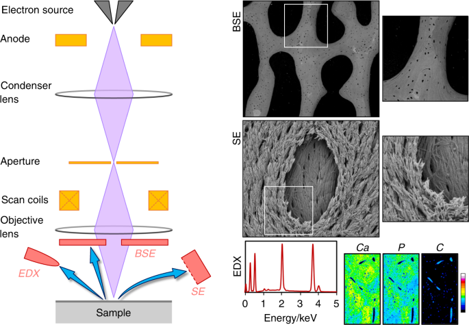
50 Years Of Scanning Electron Microscopy Of Bone A Comprehensive Overview Of The Important Discoveries Made And Insights Gained Into Bone Material Properties In Health Disease And Taphonomy Bone Research from media.springernature.com Explain the role of bone salts and the organic matrix in making bone both hard and flexible. In three dimensions an osteon is cylindrical in shape. A photo taken through a microscope that shows the anatomy of compact bone with a detailed view of an osteon. (b) in this micrograph of the osteon, you can clearly see the concentric lamellae and central canals. Do you want to learn the details of the histology of compact bone with labelled diagram and authentic slide images? This human bone section shows the haversian canal (or osteon) structure of compact bone tissue. (b) in this micrograph of the osteon, you can clearly see the concentric lamellae and central canals. Compact bone is formed in concentric circles.
The cells of compact bone, which is also called cortical bone, appear to be tightly packed into a solid mass.
3 mature bone cells, osteocytes, are found in tiny cavities within the matrix called lacunae. Compact bone is formed in concentric circles. Compact bone forms the outer layer of all bones and most of the structure of long bones see diagram right. Although the calls are close together, this type of bone is not completely solid. Learn vocabulary, terms, and more with flashcards, games, and other study tools. Compact bone forms the surface of all bones. Under the microscope, bone can be divided into two types compact bone forms the outer 'shell' of bone. Bone tissue is one of the main components of the skeletal system (other components include bone marrow/marrow cavity, collagen fibers etc). If you look at compact bone under the microscope, you will observe a highly organized arrangement of concentric circles that look like tree trunks. This video describes the microscopic anatomy of compact bone. Bone tissue and cells under the microscope introduction. Each group of concentric circles (each tree) makes up the microscopic structural unit of compact bone called an osteon (this is also called a haversian system). The marrow in these images is red marrow.
The osteon consists of a central canal called the osteonic (haversian) canal, which is surrounded by concentric rings (lamellae) of. (b) in this micrograph of the osteon, you can clearly see the concentric lamellae and central canals. Explain the role of bone salts and the organic matrix in making bone both hard and flexible. Do you want to learn the details of the histology of compact bone with labelled diagram and authentic slide images? Bone tissue is one of the main components of the skeletal system (other components include bone marrow/marrow cavity, collagen fibers etc).
Pin On A P from i.pinimg.com This video describes the microscopic anatomy of compact bone. If you look at compact bone under the microscope, you will observe a highly organized arrangement of concentric circles that look like tree trunks. Good, here in this part, i am going to describe the structure of compact bone. Compact bone, spongy if you were to look at a piece of compact bone without the help of a microscope, it would seem to be. The trabeculae are only a few cell layers thick. You can think of compact bone as being very similar. Under magnification you can clearly see the system of concentric circles that forms compact bone. Compact bone is formed in concentric circles.
The bone of the shaft of a long bone is a thick layer of compact bone.
The central canal, lamellae, canaliculi, and lacunae with osteocytes are apparent. Compact bone diagram microscope compact bone diagram parts of spongy bone diagram just wiring data structor of bone diagram wiring diagram nl the gross and microscopic structure of a long and a flat bone bones types structure and function 8 bone. The bone of the shaft of a long bone is a thick layer of compact bone. 2 compact bone we know that compact bone is very dense it is also very complex when viewed under a microscope. Compact bone is formed in concentric circles. Lamellar bone makes up the compact or cortical bone in the skeleton, such as the long bones of the legs and arms. Under magnification you can clearly see the system of concentric circles that forms compact bone. Describe the process of bone formation and bone remodeling from fetus through adulthood compact bone • dense bone Compact bone forms the outer layer of all bones and most of the structure of long bones see diagram right. If you look at compact bone under the microscope, you will observe a highly organized arrangement of concentric circles that look like tree trunks. Bone tissue and cells under the microscope introduction. Compact bone forms the surface of all bones. Explain the role of bone salts and the organic matrix in making bone both hard and flexible.
Compact bone, also called cortical bone, dense bone in which the bony matrix is solidly filled with organic ground substance and inorganic salts, leaving only tiny spaces (lacunae) that contain the osteocytes, or bone cellscompact bone makes up 80 percent of the human skeleton; compact bone diagram. Like other tissues in the body, bones are made up of specialized cells that serve different functions.


0 Komentar